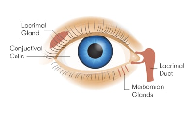
Parts of the eye
Despite their small size, your eyes contain nearly a dozen parts:
-
Sclera
-
Retina
-
Cornea
-
Lens
-
Iris
-
Pupil
-
Macula
-
Optic nerve
-
Conjunctiva
-
Aqueous humor
-
Vitreous humor
Don't know a lens from a cornea? Relax! We've got you covered. Join us on a journey through the parts of the eye.

Order Contact Lenses Online
Learn how to order contact lenses online at 1-800 Contacts
Order contacts
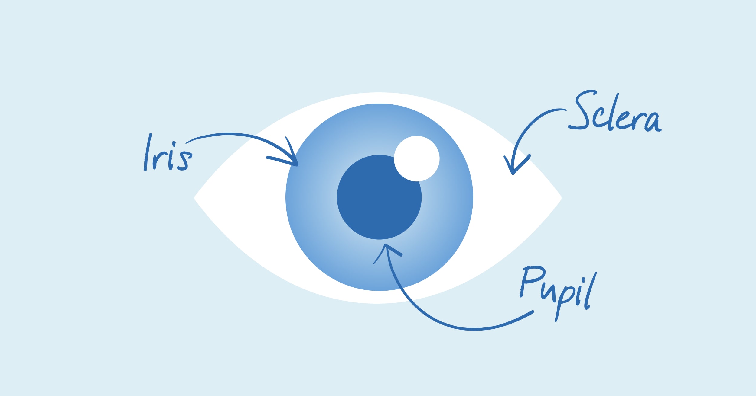
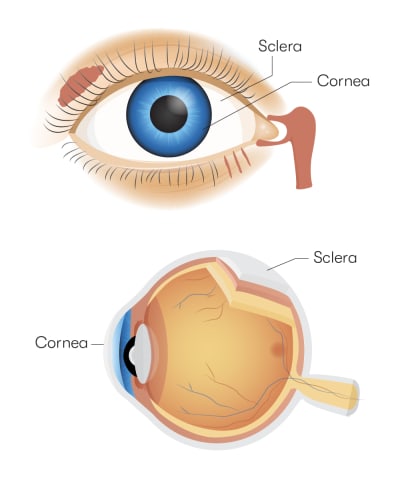
Sclera
What it is: Sclera is the technical term for the white part of your eye. It's what keeps all the squishy stuff where it belongs. Composed of collagen and elastic fibers, the sclera wraps around much of your eye, keeping the retina and the cornea (more about them later) in place.
How it works: Without sclerae, your retinas, pupils, and other parts of the eye would just be hanging there like the world's weirdest set of jewelry. The sclera is also essential for eye movement, as it's where your eye muscles attach. If you didn't have a sclera in each eye, you wouldn't be able to roll your eyes at a bad joke or wink at your latest crush.
If one of your sclerae (that's the plural form of sclera) is red and swollen, you might have a condition known as scleritis. Nodular scleritis affects just one part of the sclera, while diffuse scleritis affects the whole thing. Scleritis causes pain, increased sensitivity to light, and eye watering, so we recommend that you head to your eye care professional's office as soon as you notice any symptoms.
Retina
What it is: The retina is the thin layer of nerve cells at the back of each eye. It works with the optic nerve to turn visual stimuli (i.e., objects or movements) into images that you can see.
How it works: Surprisingly, your eyes don't actually see the things around you. Instead, they capture light and turn it into an electrical signal that travels to the brain. It's the visual cortex of the brain that "sees" everything. However, you wouldn't see anything if you didn't have retinas to detect light. Each retina also contains rods and cones. Rods help you see in low light, while cones help you detect colors.
Retinal tears and retinal detachment can both interfere with the functions of the retina. A tear occurs when the vitreous in your eye (don't worry; we'll give you the 411 on vitreous later) pulls on your retina, causing it to split open. Retinal detachment is when your retina pulls away from its supporting tissues. Both conditions can cause eye floaters or sudden flashes of light.
Cornea
What it is: The cornea has two important functions. First, it helps focus light on the retina. This is what allows the retina to convert light into signals that can travel to the visual cortex of your brain. Second, the cornea protects your eye, kind of like a helmet for your eyeball.
How it works: The cornea has six layers that work together to protect your eye. Its outermost layer, the epithelium, is incredibly thin. However, it does an excellent job keeping dirt and debris away from your eye. Bowman's layer contains collagen, a protein that helps tissue maintain its structure. Collagen helps your cornea maintain its natural shape.
The stroma does most of the heavy lifting when it comes to bending light. It's the thickest layer of the cornea, so it's extra durable. The other three layers help maintain your eye's structure, achieve the right balance of fluid in the eye, and separate the fluid in your eye from the air you breathe.
Dry eye and keratitis are two of the most common corneal conditions. Have you ever spent the day rubbing your eyes furiously? Feeling like you were wandering through the desert without a drop of moisture in sight? You might have dry eye, a lack of tear fluid around the epithelium.
Keratitis is just a fancy way of saying corneal inflammation. It can cause eye redness, pain, blurry vision, excessive tearing, and sensitivity to light. You may also feel like you have something stuck in your eye.
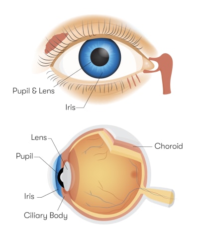
Lens
What it is: The lens is the structure of the eye that focuses light and directs it to the retina. It's also known as the crystalline lens. This name comes from the proteins — crystallins — that give it its structure.
How it works: When light enters your eyes by way of your pupils, the lenses pull triple duty. They absorb the light, focus it and send it to the retina.
Cataracts are the most common disorder affecting the lens of the eye. A cataract forms when the proteins in your eye start to break down. This causes the lens to become cloudy, making it difficult to see clearly. It's like trying to drive a car through a dust storm. Sure, you might be able to make it safely, but it's far from easy.
Iris
What it is: If the parts of the eye were a baseball team, the iris would be a great utility player. It's what gives your eye its color, but it also contains the muscles that allow your pupils to get bigger or smaller.
How it works: The iris controls how much light enters your eye. If you're outside on a sunny day, it helps your pupil contract (get smaller). In a low-light environment, the iris helps your pupil dilate (get bigger).
The irises are pretty low maintenance, but some people develop a condition known as iritis. This is when the iris becomes inflamed, causing eye pain, headaches, light sensitivity, and vision changes.
Pupil
What it is: The pupil is the black part of the eye. It works with the iris to control the amount of light entering your eye at any given moment.
How it works: The pupil is kind of like the aperture of a camera lens. In a camera, the aperture determines how much light hits the image sensor. The pupil expands and contracts based on changes in your environment. If you step outside after spending time in a dark room, your pupils will contract to prevent too much light from getting in. Conversely, they'll expand if you walk from a bright environment into a dark one.
In people with physiologic anisocoria, the pupils are slightly different sizes. Uneven pupils can also occur due to brain aneurysms or torn blood vessels in the neck.
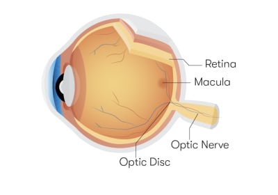
Macula
What it is: The macula is a tiny part of your retina. It's responsible for central vision, which is the ability to see objects directly in front of you.
How it works: Each macula has tons of photoreceptor cells. In simple terms, photoreceptor cells receive light. Once the cornea and the lens help focus light on the retina, the macula converts light into specialized signals. These signals travel to the brain, transforming into images you can see — almost like magic.
Macular degeneration is one of the most common conditions affecting this part of the eye. The dry form occurs when protein deposits form under the macula. This causes the macula to get thin and dry, like an old corn husk. Wet MD develops when blood vessels under the macula and retina start to leak fluid and blood. This causes the macula to bulge.
Optic Nerve
What it is: The optic nerve transports information from your retinas to your brain, allowing you to see the world around you.
How it works: The optic nerve contains millions of nerve fibers, so it's the MVP of carrying electrical signals to your visual cortex. Some of these fibers also help your pupils adjust to different levels of light. The optic nerve is even responsible for maintaining your Circadian rhythm, or the internal clock that controls your sleep-wake cycle.
Optic neuritis develops when one of the optic nerves becomes inflamed. As you can imagine, it's a pretty painful condition. It can also reduce the sharpness of your vision or make it difficult to see colors clearly.
Conjunctiva
What it is: The conjunctiva is the thin layer of tissue that covers the sclera. It's one of your eye's biggest defenders, as it produces tears and mucus. It also prevents bacteria from getting deeper into your eye, so it's like an infection-fighting superhero.
How it works: This protective structure has three parts:
-
Palpebra conjunctiva: Lines your eyelids
-
Bulbar conjunctiva: Covers your eyeball
-
Conjunctiva fornix: Connects the bulbar conjunctiva with the palpebra conjunctiva
All three parts work together to keep moisture in and keep bad things out.
Conjunctivitis is one of the most common conditions of the human eye. Better known as pink eye, conjunctivitis occurs due to inflammation. It can develop due to the presence of bacteria or viruses, but it's also a symptom of environmental allergies.
Aqueous humor
What it is: Aqueous humor is the fluid that fills the space behind your corneas.
How it works: This clear fluid helps your eye maintain its natural shape. It also nourishes the eye, which helps to keep it healthy. No matter what time of day it is, your eyes are producing new aqueous fluid and allowing an equal amount of fluid to drain into your bloodstream.
If your eyes make too much aqueous humor, or they don't drain enough aqueous humor, you might develop a condition known as glaucoma. This condition causes the pressure in your eye to increase, which can damage your vision.
Vitreous humor
What it is: Next up in our eyeball anatomy tour is the vitreous humor, a clear, gel-like fluid that hangs out between the lens and the retina.
How it works: Like aqueous humor, vitreous humor helps your eye maintain its natural shape. It's attached to the retina with small fibers.
As you age, these fibers can break away from the retina, resulting in a condition called vitreous detachment. If you have vitreous detachment, you might notice floaters or have problems with your peripheral vision (what you see to your side as you're looking straight ahead).
What is the stuff in the corner of your eye called?
That gunk in the corner of your eye is called eye discharge. Sounds gross, but it's actually a good thing. Day and night, your eye is cleaning itself with tears and mucus. When you blink, eye discharge disappears into the ether. At night, however, you don't blink as much, so you might wake up with crust in the corner of each eye. No worries. Simply wipe it away when you're ready to start your day.
If your eye is irritated or infected, it might produce too much discharge. Book an appointment with an eye care professional if you're concerned.
Human eye diagram
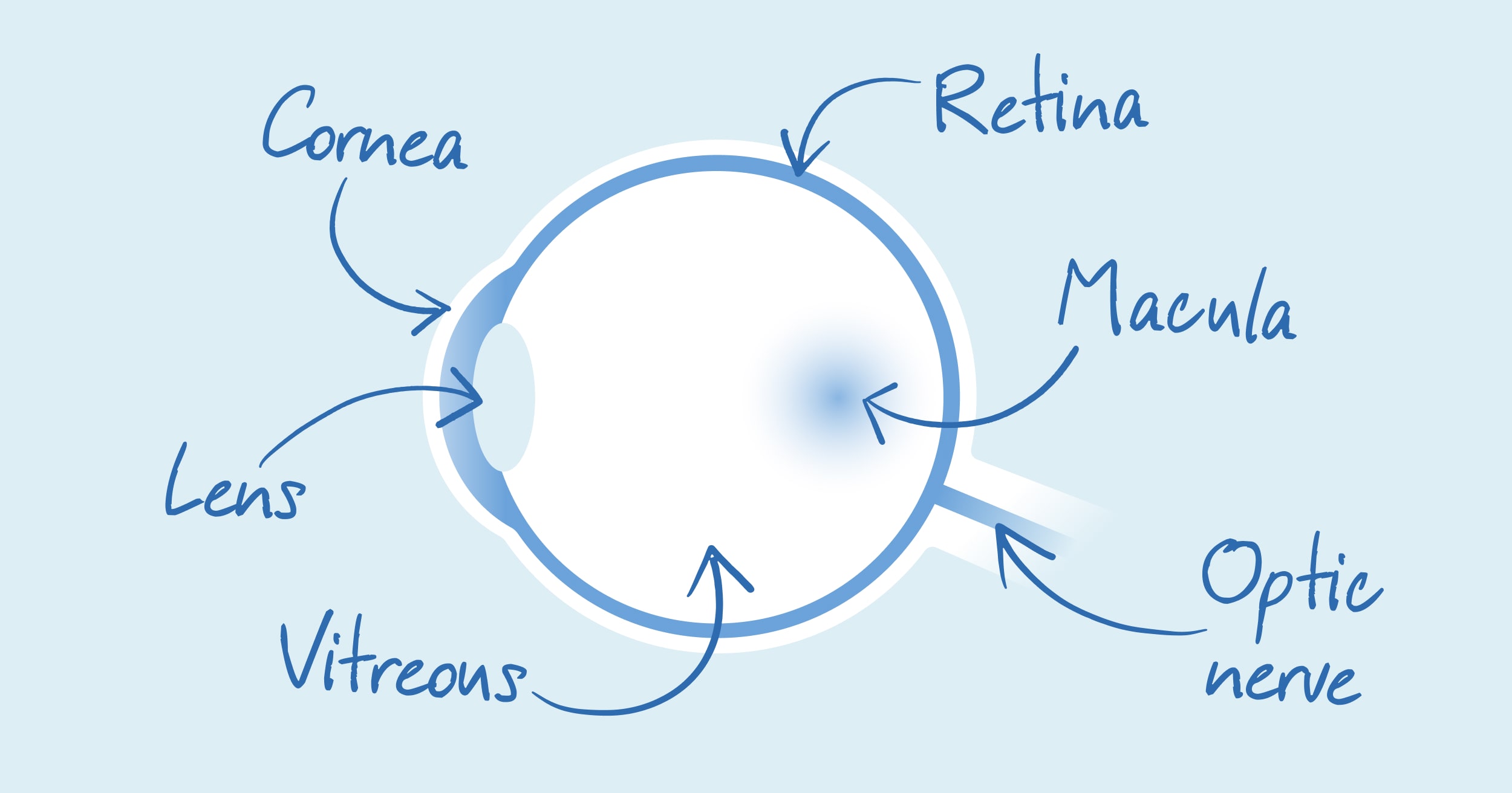
Structure of the eye
Now that you understand the anatomy of the eye, here's an overview of how all the parts are arranged:
-
Cornea. The cornea is at the very front of the eye. Think of it as the welcome wagon for all the light around you.
-
Aqueous humor. Aqueous humor hangs out just behind the cornea.
-
Iris. Your irises sit behind the aqueous humor.
-
Pupil. Your pupils are the black spots inside your irises. They're like best friends trapped in an eyeball.
-
Lens. The lens sits behind the pupil.
-
Vitreous humor. Next comes the vitreous humor. It sits between the lens and the retina.
-
Retina. Now we're in the back of the eye. You can't see it, but it's always working double-time to turn light into electrical signals.
-
Macula. The macula is part of your retina, so it's also behind the vitreous humor.
- Optic nerve. Last but not least, the optic nerve is at the veeeeery back of your eye.

Online Vision Exam at 1-800 Contacts
Renew your prescription online with ExpressExam
Try ExpressExam
Eye anatomy
There you have it. All the eye parts, their names, and almost everything you need to know about them. If you have floaters, eye pain, or other symptoms, consult an eye care professional for guidance. In the meantime, use our website to find your preferred brand of contact lenses or schedule an online eye exam.
Did you know you can order contacts online?
Say goodbye to trips to the eye doctor and hello to convenience! With 1-800 Contacts, you can easily order your contact lenses online and have them delivered straight to your door. Plus, our online vision exam makes it simple to get the prescription you need without leaving home. And the best part? You’ll save money on your first order with 1-800 Contacts. It’s never been easier to get your contacts!
Original published date: 5/13/2020
Updated: 9/8/2025